




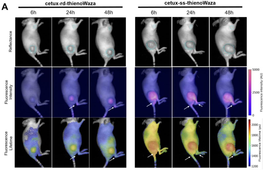
Representative tauCAM in vivo reflectance, fluorescence intensity and FLT images of mice bearing a EGFR+ subcutaneous tumor (right lateral view) acquired at 6, 24 and 48h after intravenous administration of cetux-rd-thienoWaza or cetux-ssthienoWaza. On the reflectance images, ROIs drawn around the tumor (T), the tumor margin (TM) and the shoulder (S) (background normal tissue) are indicated. Sites with non-specific fluorescent signals on the legs and tail basis are indicated with arrows.
